Diplozoon paradoxum
Geographic Range
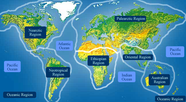
Diplozoon paradoxum is the parasite of various cyprinid fishes in Europe (and possibly Asia) (Schmidt & Roberts, 1989). (Schmidt and Roberts, 1989)
- Biogeographic Regions
- palearctic
- oriental
Habitat
Diplozoon paradoxum individuals parasitize various cyprinid fishes, including, but not limited to: Abramis, Rutilus, Gobio, and Phoxinus. A study in Northern England found that Diplozoon paradoxum were absent entirely from fish of the genus Leuciscus (Wiles, 1968). (Wiles, 1968)
- Habitat Regions
- freshwater
- Other Habitat Features
- agricultural
Physical Description
An adult specimen of Diplozoon paradoxum is two permanently fused individuals, conjoined at the flat fusion bridge, so that their bodies form an H shape together (Wong & Gorb, 2013). Adult Diplozoon paradoxum were measured at 8 to 10 mm in length (Schmahl & Taraschewski, 1987), and the fusion bridge is 0.37 mm in length (Wong & Gorb, 2013). The larva, or diporpa, reported by Bychowsky (1957) are roughly 0.23 mm in length. (Bychowsky, 1957; Schmahl and Tarachewski, 1987; Wong and Gorb, 2013)
The integument of Diplozoon paradoxum is comparatively irregular in comparison to closely related species, likely attributable to many vacuoles and vesicles. It is a syncytium, measuring roughly 2 to 6 um deep, with mitochondria often lying near the base (Schmahl and Tarachewski, 1987). (Schmahl and Tarachewski, 1987)
The individuals of the mated pair each maintain their own anterior attachment apparatus, consisting of two buccal suckers lateral to the mouth. These suckers are aided by groupings of gland cells that secret an adhesive substance. The mouth is located on the anterior ventral surface of the parasite, it is followed by a buccal cavity, where the oval shaped pharynx is slightly protruded (Halton & Jennings, 1965). The species is also in possession of a comparatively long trapeze spur (Matejusova et al., 2004). (Halton and Jennings, 1965; Matejusová, et al., 2004)
The opisthaptor of adults of this species, uses four pairs of clamps that are arranged in parallel lateral rows on the ventral posterior terminal end of the body. There is also a small pair of hooks on the ventral side of the haptor, though the clamps are thought to play the most crucial role in attachment to the substrate. The clamps are supported by a framework of sclerites, and receive peripheral support by marginal sclerites. The anterior end of the clamps is fixed, and the posterior end is hinged. The sclerites of the clamps are covered in a fibrous tissue layer, that is roughly 0.01 mm thick (Owen, 1963). Recent studies have shown the clamps remain closed by nature of the elastic material of the clamps, not active muscular force. It is believed that the clamps are only opened by muscle force, which is a likely explanation of the long term fixed position of adult specimens. There is a positive relationship between the force required for removal from host tissue and the body mass of the parasite. The force to mass ratio is roughly 246, which is considered high among other studied invertebrate species (Wong & Gorb, 2013). Each clamp is able to grasp one or two gill lamellae, though the first, most posterior clamp is sometimes smaller than the other three (Wong & Gorb, 2013). All clamps of the opisthaptor of D. paradoxum were of the same size (Matejusova et al., 2004). In another study concerning abnormalities of the attachment clamps, the precedent was to consider any clamps of a smaller size, abnormal (Sebelova et al., 2002). The clamps are innervated by two nerves each, that both bifurcate, and travel one branch into the posterior, and one branch into the anterior portion of the clamp, where on both side they branch excessively, and end at the distal tip of the tissue lining (Owen, 1963). It is also noteworthy, that all eight clamps were observed by Wong and Gorb (2013) to be able to open and close independent of each other. (Matejusová, et al., 2004; Owen, 1963; Šebelová, et al., 2002; Wong and Gorb, 2013)
The excretory system of D. paradoxum is comprised of two different types of cells, flame cells and canal cells. The flame cells are flagellated and the canal cells line the anterolaterally-placed excretory ducts. The collecting ducts are interwoven throughout the body, and are greatest in number in the anterior and posterior terminal regions. The portions of the canal cells lining the excretory ducts are ciliated to assist in the transportation of liquid through the canal (Konstanzova et al., 2016). (Konstanzová, et al., 2016)
- Other Physical Features
- ectothermic
- bilateral symmetry
-
- Range length
- 8 to 10 mm
- 0.31 to 0.39 in
Development
The eggs of the oviparous parasites have long coiled filaments at their ends (Schmidt & Roberts 1989), and can hatch nearly ten days after deposition (Bychowsky, 1957). Light intensity and turbulence of water, which might be caused by host spawning, or feeding activity, assist in stimulating hatching (Bychowsky, 1957). A newly-emerged oncomiracidium is ciliated, possesses two pair of clamps on the opisthaptor, and bears two medial hooks that are quite large, and assist in both the movement and the settlement of the larva (Bychowsky, 1957). The cilia are localized in three places on the body by five different groups of cells. The first group of cilia are on the lateral and dorsal surfaces of the body, anterior to the small band, but not on the anterior end of the organism. The second grouping of cilia, comprised of two sets of cells, begins posterior to the small band, and continues on the lateral and dorsal surfaces, until reaching the anterior end of the opisthaptor. The third grouping of cilia is behind the posterior attachment apparatus on the lateral and dorsal surfaces of the animal, and continues posteriorly to the terminal end of the animal. (Bychowsky, 1957; Llewellyn, 1963). Bychowsky (1957) identified two fundamental phases of larval activity, a free swimming phase, making use of the cilia, and a somewhat attached phase where the larva creeps through the substrate using its musculature. Attachment to the host occurs early in diporpa stages of development (larval stages specific to this clade of parasites; Schmidt & Roberts, 1989), often utilizing only the buccal suckers (Kagel & Tarachewski, 1993). After attaching to a gill filament, the larva loses its cilia almost immediately. The worm will feed and grow, acquiring another set of clamps, a dorsal papilla, and a ventral sucker, posterior to the buccal sucker. The growth and development of the clamps occurs in order, with the first pair at the most posterior end of the opisthaptor to the fourth pair at the most anterior end (Sebelova et al., 2002). The growth of the third set of posterior clamps in one individual can be asymmetrical, with one side of the opisthaptor having two clamps, and the other side having three (Bychowsky, 1957). Structural abnormalities such as the opisthaptoral clamps can be traced to deviations in normal development, often from the embryonic state (Sebelova et al., 2002). Upon mating the pigmented eyes disappear, and as the larva grow into each other, and merge, they acquire their final sets of clamps and grow to their full size. Growth of the individual occurs rather slowly, and accelerates greatly after mating. If independent larvae do not mate they perish by the following winter. Mated pairs undergo rapid growth, and reach maturity by spring of the following year (Bychowsky, 1957). (Bychowsky, 1957; Kagel and Tarachewski, 1993; Llewellyn, 1963; Schmidt and Roberts, 1989; Šebelová, et al., 2002)
- Development - Life Cycle
- metamorphosis
Reproduction
Diplozoon paradoxum is oviparous, and production of eggs is only achieved by paired, fully matured organisms (Koskova et al., 2011; Bychowsky, 1957). Two hermaphroditic individuals must meet and permanently fuse their bodies in order to achieve maturity (Avenant-Oldewage & Milne, 2014). Egg production occurs at its highest rates in the warm months, May through June, but does continue through the summer (Bychowsky, 1957). Virtually no gametes are produced during the winter (Schmidt & Roberts, 1989). According to Zeller (1872, as cited in Bychowsky 1957), the rapid development of the egg cells and vitelline cells takes place in the spring, along with the concurrent rise in temperature. An artificial introduction of the parasite and its host, to warmer temperatures saw egg formation begin about the fifth or sixth day. The vitelline glands are in close contact with the intestine for a majority its length (Halton & Jennings, 1965). Diporpa larva each develop a dorsal papilla and a ventral sucker, posterior to the buccal cavity. When the worms mate, one individual attaches its ventral sucker to the papilla on the dorsal surface of the other. This encounter stimulates fusion, maturation, and the gonads finally complete development (Schmidt & Roberts, 1989). According to Bovet (1967, as cited by Anderson 1974), once the hermaphroditic larvae mate, the vagina of one individual is opened in close proximity of the vas deferens of the other, resulting in cross fertilization between the adult, fused, mated pair. (Avenant-Oldewage and Milne, 2014; Bychowsky, 1957; Halton and Jennings, 1965; Košková, et al., 2011; Schmidt and Roberts, 1989; Zeller, 1872)
- Key Reproductive Features
- simultaneous hermaphrodite
- oviparous
- Parental Investment
- no parental involvement
Lifespan/Longevity
The infective stages of Diplozoon paradoxum have a lifespan of approximately 10 hours (Bovet, 1967, as cited in Anderson 1974). The diporpa stage of the species is capable of living for several months, once attached to a host, but without a mate, will die in the winter months. Mated pairs are able to live off of the host for several years (Schmidt & Roberts, 1989). (Anderson, 1974; Bovet, 1967; Schmidt and Roberts, 1989)
Behavior
A study of the common bream (Abramis brama) found that the number of parasites per host increases with the age of the fish, and that parasite burden is determined by three factors. First, Bovet (1967) suggests that adult specimens anchor themselves permanently to their respective hosts. Because of this permanent settlement, a single organism claims a defined space on the gill lamellae. The parasites compete for space on the gill filaments, and by association, the blood supply from which they obtain nutrients. Overcrowding of this environment will often result in failure of an organism to find a mate, hindering maturation and survival capability, or failure of the organism to attach to the substrate. Second, older fish are bound to accumulate more parasites over time, resulting in a larger burden. Third, a larger fish passes a larger volume of water over their gills. If this water is infected, then larger fish have higher rates of exposure to infection (Anderson, 1974). Above average desnities of D. paradoxum have detrimental effects on the mass of the host in the winter and spring months, potentially resulting in the death of the host. This occurs especially where specific age groups of fish compete for limited resources. Different developmental stages of the parasite have different microhabitat preferences. Diporpa are most frequently found on the distal portion of the primary gill lamellae, and often only attached to the host by their anterior end. Usually unpaired diporpa only enclose one secondary gill lamellae with the underdeveloped opisthaptor, and are much less restricted in their mobility. Adult individuals have a preference for the basal portion of the primary gill lamellae, however, and each clamp is capable of compressing between one and four secondary gill lamellae (Kagel & Tarachewski, 1993). A more recent study found the monogeneans localized at the gill filaments “tightly adhering to the gills by the opisthaptor while also concentrating in small groups, with no preferences in localization between hemibranches or gill arches" (Yildirum & Uvner, 2012:246). Wiles (1968) found that infection rates of Diplozoon paradoxum were highest in moving bodies of water, and lowest in stagnant water such as ponds and reservoirs. Diplozoon paradoxum is known to infect more than 30 species of cyprinid fish and several species of Cobitidae. Bychowsky (1957) asserts that they do not parasitize any fish species except representatives from these two families. (Anderson, 1974; Bovet, 1967; Bychowsky, 1957; Kagel and Tarachewski, 1993; Wiles, 1968; Yıldırım and Üvner, 2012)
- Key Behaviors
- parasite
Communication and Perception
The diporpa phase of the species is in possession of four eyes, each having a single rhabdomere. Two of these eyes are laterally arranged on the anterior midline, near the dorsal surface, and covered by a pigment shield. They are cup shaped and are oriented laterally. These eyes have no solid lenses and no oil droplets that might otherwise serve as temporary lenses. A single retinular cell is associated with each of these pigmented eyes. Muscle fibers are abundant in the vicinity of the eyes, but direct articulation between those structures has not been confirmed. The eyes lacking pigment shields lie lateral to the pharynx, and closer to the surface of the body. They too are rhabdomereic and closely resemble the other pair of eyes, apart from lacking the pigment shield. No apparent movement has been observed in these eyes, but it is hypothesized that they are useful in allowing the animal to orient itself. It is likely that the lateral eyes are well equipped to detect sudden changes in light intensity, and this can be related to the perception of the shadow casting of a fish (Kearn, 1978). (Kearn, 1978)
- Perception Channels
- visual
Food Habits
These sanguivorous parasites feed using negative pressure to suck a plug of gill tissue into the buccal cavity back to the pharynx, where prolonged strain on the tissue eventually causes it to rupture. This rupture is the result of negative pressure alone, and is not aided by any glandular secretions. The ruptured tissue permits the low of blood, and some tissue fragments This sanguinivorous diet is relatively characteristic of the polyopisthocotylea, however Diplozoon paradoxum was found to supplement the blood diet with variable amounts of gill tissue and mucus. Hemolysis occurs almost immediately within the buccal cavity, and is followed by "partial intraluminar digestion" within the posterior, laterally-branched intestine. This intestine extends, in each individual, to the fusion bridge, where the intestines of the two organisms merge. In the intestine, "soluble substances are absorbed by the gastrodermis," (Halton & Jennings, 1965:266) and subsequently the remainder of digestion occurs in the intracellular space. Once the erythrocytes have been stripped of the useful byproducts, hematin is left in the intracellular space, which requires that the organism disintegrate older cells. This process likely results in the branched nature of the intestine, and indicates an incomplete evolution of the parasites toward the blood diet. A positive side effect is that this destruction of gastrodermal cells releases intracellular enzymes that aid in extracellular digestion (Halton & Jennings, 1965). (Halton and Jennings, 1965)
- Primary Diet
-
carnivore
- sanguivore
- eats body fluids
- Animal Foods
- blood
- body fluids
Predation
No information exists at this time
Ecosystem Roles
- Ecosystem Impact
- parasite
- carps and true minnows (Cyprinidae)
Economic Importance for Humans: Negative
Gill parasitizing monogeneans often become a serious problem in commercial fish farms, and have been a serious issue in Eastern and southeastern Europe. First noticed in the early 1950s, the pathological effects of these parasites can result in lethal damage to the host. Many treatments for monogeniasis, can be highly toxic for the fish as well. Praziquantel is the most effective means of killing monogenean parasites, without adverse effects to the host (Schmahl & Taraschewski, 1987). (Schmahl and Tarachewski, 1987)
Other Comments
Although generally visible in the natural coloring of a specimen, Halton and Jennings (1964) found a strong reaction of the nervous system to non-specific esterase, and this reaction was specific to Diplozoon paradoxum; it was observed minimally in other species of monogeneans. Koskova et al. (2011) provided a phylogenetic tree based on RNA ITS, which is corroborated by karyological analysis (Matejusova et al., 2004). (Halton and Jennings, 1964; Košková, et al., 2011; Matejusová, et al., 2004)
Contributors
Steve Mullin (editor), Stephen F. Austin State University, Tanya Dewey (editor), University of Michigan-Ann Arbor.
Glossary
- Palearctic
-
living in the northern part of the Old World. In otherwords, Europe and Asia and northern Africa.
- agricultural
-
living in landscapes dominated by human agriculture.
- benthic
-
Referring to an animal that lives on or near the bottom of a body of water. Also an aquatic biome consisting of the ocean bottom below the pelagic and coastal zones. Bottom habitats in the very deepest oceans (below 9000 m) are sometimes referred to as the abyssal zone. see also oceanic vent.
- bilateral symmetry
-
having body symmetry such that the animal can be divided in one plane into two mirror-image halves. Animals with bilateral symmetry have dorsal and ventral sides, as well as anterior and posterior ends. Synapomorphy of the Bilateria.
- bog
-
a wetland area rich in accumulated plant material and with acidic soils surrounding a body of open water. Bogs have a flora dominated by sedges, heaths, and sphagnum.
- carnivore
-
an animal that mainly eats meat
- ectothermic
-
animals which must use heat acquired from the environment and behavioral adaptations to regulate body temperature
- freshwater
-
mainly lives in water that is not salty.
- marsh
-
marshes are wetland areas often dominated by grasses and reeds.
- metamorphosis
-
A large change in the shape or structure of an animal that happens as the animal grows. In insects, "incomplete metamorphosis" is when young animals are similar to adults and change gradually into the adult form, and "complete metamorphosis" is when there is a profound change between larval and adult forms. Butterflies have complete metamorphosis, grasshoppers have incomplete metamorphosis.
- native range
-
the area in which the animal is naturally found, the region in which it is endemic.
- oriental
-
found in the oriental region of the world. In other words, India and southeast Asia.
- oviparous
-
reproduction in which eggs are released by the female; development of offspring occurs outside the mother's body.
- parasite
-
an organism that obtains nutrients from other organisms in a harmful way that doesn't cause immediate death
- pelagic
-
An aquatic biome consisting of the open ocean, far from land, does not include sea bottom (benthic zone).
- sanguivore
-
an animal that mainly eats blood
- swamp
-
a wetland area that may be permanently or intermittently covered in water, often dominated by woody vegetation.
- visual
-
uses sight to communicate
References
Anderson, R. 1974. An analysis of the influence of host morphometric features on the population dynamics of Diplozoon paradoxum (Nordmann, 1832). Journal of Animal Ecology, 43/3: 873-887.
Avenant-Oldewage, A., S. Milne. 2014. Aspects of the morphology of the juvenile life stages of Paradiplozoon ichthyoxanthon Avenant-Oldewage, 2013 (Monogenea: Diplozoidae). Acta Parasitologica, 59/2: 247-257.
Bovet, J. 1967. Contribution à la morphologie et à la biologie de Diplozoon Paradoxum v. Nordmann, 1832. Bull. Soc. Neûchatel Sci. nat., 90: 64-159.
Bychowsky, B. 1957. Monogenetic Trematodes: Their Systematics and Phylogeny. Washington D.C.: American Institute of Biological Sciences.
Halton, D., J. Jennings. 1964. Demonstration of the nervous system in the monogenetic trematode Diplozoon paradoxum Nordmann by the indoxyl acetate method for esterases. Nature, 202/4931: 510-511.
Halton, D., J. Jennings. 1965. Observations on the nutrition of monogenetic trematodes. Biological bulletin, 129/2: 257-272.
Kagel, M., H. Tarachewski. 1993. Host-parasite interface of Diplozoon paradoxum (Monogenea) in naturally infected bream, Abramis brama (L.). Jornal of Fish Disease, 16/5: 501-506.
Kearn, G. 1978. Eyes with, and without, pigment shields in the oncomiracidium of the monogenean parasite Diplozoon paradoxum. Zeitschrift für Parasitenkunde, 57/1: 35-47.
Konstanzová, V., B. Koubková, M. Kašný, J. Ilgová, E. Dzika, M. Gelnar. 2016. Excretory system of representatives from family Diplozoidae (Monogenea). Parasitology Research, 115/4: 1493-1500.
Košková, E., M. Špakulová, B. Koubková, M. Reblánová, M. Orosová. 2011. Comparative karyological analysis of four diplozoid species (Monogenea, Diplozoidae), gill parasites of cyprinid fishes. Parasitology Research, 108/4: 935-941.
Llewellyn, J. 1963. Larvae and larval development of mongeneans. Advances in Parasitology, 1: 287-326.
Matejusová, I., B. Koubková, C. Cunningham. 2004. Identification of European diplozoids(Monogenea, Diplozoinae) by restriction digestion of the ribosomal RNA internal transcribed spacer. Journal of Parasitology, 90/4: 817-822.
Owen, I. 1963. The attachment of the monogenean Diplozoon paradoxum to the gills of Rutilus rutilus L. II. Structure and mechanism of the adhesive apparatus. Parasitology, 53/3-4: 463-468.
Schmahl, G., H. Tarachewski. 1987. Treatment of fish parasites. Avenant-Oldewage, 2013 (Monogenea: Diplozoidae). Parasitology Research, 73/4: 341-351.
Schmidt, G., L. Roberts. 1989. Foundations of Parasitology. St. Louis Missouri: Times Mirror/Mosby.
Wiles, M. 1968. The occurrence of Diplozoon paradoxum Nordmann, 1832 (Trematoda: Monogenea) in certain waters of northern England and its distribution on the gills of certain Cyprinidae. Parasitology, 58/1: 61-70.
Wong, W., S. Gorb. 2013. Attachment ability of a clamp-bearing fish parasite, Diplozoon paradoxum (Monogenea), on gills of the common bream, Abramis brama. Journal of Experimental Biology, 216/16: 3008-3014.
Yıldırım, M., B. Üvner. 2012. Metazoan parasites of Alburnus chalcoides in Tödürge Lake (Zara/Sivas, Turkey). Journal of Applied Ichthology, 28/2: 245-248.
Zeller, E. 1872. Untersuchungen über die Entwicklung des Diplozoon paradoxum. Leipzig Germany: Engelmann.
Šebelová, Š., B. Kuperman, M. Gelnar. 2002. Abnormalities of the attachment clamps of representatives of the Family Diplozoidae. Journal of Helminthology, 76/3: 249-259.