Fasciola gigantica
Geographic Range
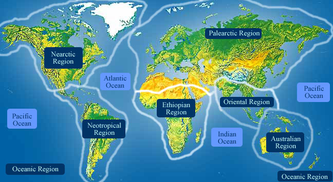
Fasciola gigantica is found in tropical Africa, South and South-east Asia, and the Far East. In the United States F. gigantica is found in Hawaii. ("Fascioliasis", 2009; Cheesbrough, 2005; Saleha, 1991)
Habitat
The habitat of Fasciola gigantica changes with the stage of its life cycle. Adult F. gigantica live in the liver and bile ducts of its definitive hosts (sheep, cattle, and other grazing ruminant mammals). Eggs shed by adults are located in the intestinal track of mammals and also in the wild. Free swimming cercarias are found in the bodies of fresh water that are in close proximity of its definitive host. Temperatures above 10 degree Celsius are required for the development of the miracidia larvae stage. Miracidia are found in fresh water that contains intermediate snail hosts in the genus Lymnaea. Metacercaria are found encysted on vegetation and in the mammal host once ingested. Inside the snail rediae persist in the digestive gland of the mammal host, known as the hepatopancreas.
Adequate amounts of moisture are also needed, these factors can account for the intensity and prevalence of infection in the definitive hosts. Increased numbers of incidences are seen in the wet season. (Cheesbrough, 2005; Read, 1973)
- Habitat Regions
- tropical
- freshwater
- Aquatic Biomes
- lakes and ponds
Physical Description
Fasciola gigantica is leaf-shaped and tapers at both ends. An adult can grow to 75 mm in length. With the use of a scanning electron microscope the surface of F. gigantica appears very rough due to abundant microscopic spines and surface folding. Spines increase in size in their middle section and are smaller on the surface near the suckers. Spikes range from 30 μm to 58 μm and have serrated edges with anywhere from 16 to 20 sharp points. This species has both an oral and ventral sucker for feeding and attaching to the inside of its host. Fasciola gigantica also has three different types of surface papillae which are used as sensory receptors. The eggs of F. gigantica can reach sizes of 0.2 mm in length. (Dangprasert, et al., 2001; Kumar, 1998)
- Other Physical Features
- ectothermic
- heterothermic
- bilateral symmetry
- Sexual Dimorphism
- sexes alike
-
- Range length
- 25 to 75 mm
- 0.98 to 2.95 in
Development
Adult flukes produce eggs that are passed in the host’s feces. In the wild the eggs hatch into miracidia and penetrate a snail host. The miracidia form into saclike sporocysts and multiply into rediae which then develop into cercariae. Free swimming cercariae are shed from snails, they then find aquatic plants, encyst and become metacercariae. Metacercariae cysts wait to be taken up by other ruminant host to repeat the life cycle. In the transmission stage the metacercariae are unknowingly ingested with aquatic plants by humans and grazing mammals. In the mammal host metacercariae excyst in the duodenum. Immature flukes then penetrate the liver and become mature in the biliary track. (Cheesbrough, 2005; Read, 1973)
Reproduction
Sexually mature adults reside and presumably mate in biliary ducts of their mammalian host. ("Fascioliasis", 2009)
Fasciola gigantica reproduce sexually as adults and asexually in the other stages of its life cycle. The flukes are in the metacercariae stage before becoming sexual adults. After residing in their mammal host’s duodenum, the metacercariae penetrate the liver and become mature in the biliary track. The adult flukes have both sex organs, but fertilization between adult male and female flukes is the most common source of sexual reproduction. Adult flukes produce eggs that are then passed in the host’s feces. (Cheesbrough, 2005; Read, 1973)
- Key Reproductive Features
- sexual
- asexual
- fertilization
Non-embryonic eggs are laid within the mammalian host and are passed through to the intestinal tract where they are expelled in the feces. ("Fascioliasis", 2009)
- Parental Investment
- no parental involvement
Lifespan/Longevity
The life span for each stage of Fasciola gigantica varies greatly. After being ingested it takes 3-4 month for adult flukes to become mature and begin producing eggs. Adult flukes can live for multiple years in their definitive host. Embryos of Fasciola species are able to persist outside the host for several months. The free-swimming miracidium will die soon after hatching if they do not contact a secondary host. If conditions are favorable metacercaria are able to persist for up to a year once encysted. (Miliotis and Bier, 2003; Read, 1973)
Behavior
Adult Fasciola species produce embryos which are then shed through the digestive system with its host’s feces. When water and temperature conditions are favorable, the embryos develop into the ciliated larva form called, miracidium. Miracidium are able to swim and locate its secondary snail host with help of its cilia. If the miracidium are to come in contact with a snail it actively penetrates it. The cilia are then shed and it is transformed in to a saclike sporocyst. The sporocyst mature and release rediae. Inside the snail the rediae move to the snail’s digestive gland known as the hepatopancreas. Over time this is where the rediae will form cercariae. The cercariae develop tails and leave the snail in search for vegetation to encyst. Once the cysts are formed Fasciola species become metacercaria and these infect a mammal host if they are accidentally ingested with the vegetation. (Read, 1973)
Communication and Perception
The time it takes for the shelled embryos of Fasciola gigantica to hatch is rapidly increased in the presence of light. If they are kept in darkness the number of miracidia that make it to the free-swimming stage are greatly reduced. The miracidium have extremely functional eye-spots. This is probably to prevent premature hatching before the embryos have exited the host’s digestive track. The attack of a proteoplytic enzyme protein controls the opening of the operculum which allows the miracidium to exit the shelled embryo. There is evidence that the blue and violet portion of the light spectrum triggers this attack. (Read, 1973)
- Communication Channels
- tactile
Food Habits
When the adult Fasciola are in the bile ducts of a host it obtains a small portion of its nutrients from active bloodsucking. In a day’s time, a single adult fluke can take in about 0.2 ml of blood. There is evidence that adult flukes need around 100 times the amount of glucose than Fasciola receives from active ingestion. Therefore, adult flukes also receive nutrients by absorption of glucose through their tegument. The free-swimming miracidia were once thought to be a non-feeding stage, but it has been shown they metabolize glucose when it is present. (Read, 1973)
- Primary Diet
- carnivore
- Animal Foods
- mammals
- blood
Predation
A variety of general aquatic predators are known to feed on the free-living stages of Fasciola gigantica, including the miracidia and the cercariae stages. (Johnson and Thieltges, 2009)
Ecosystem Roles
Fasciola spp. have a negative impact on its definitive host and are capable of causing mortality if infection is severe. Fasciola gigantica parasitizes lymnaeid snails, which are intermediate hosts, and cows, sheep and other ruminants, which are definitive hosts. In some instances humans are also infected. (Miliotis and Bier, 2003)
- Ecosystem Impact
- parasite
- lymnaeid snails, Lymnaea
- sheep, Caprinae
- cows, Bovinae
- humans, Homo sapiens
Economic Importance for Humans: Positive
These are no known positive effects of Fasciola gigantica on humans.
Economic Importance for Humans: Negative
Fasciola spp. cause a disease known as fascioliasis, which can cause significant economic loss when it infects livestock. This parasite reduces the amount of meat that a cow will produce when slaughtered, and leaves the liver inedible. This parasite also reduces milk production and can causes mortality in chronic infections. In some developing countries fascioliasis is the second leading cause of economical loss, costing 10 million dollars annually. Immunization of livestock also adds to the cost of fascioliasis. Few human infections with F. gigantica have been reported but are seen in Asia, Africa, and Hawaii. ("Fascioliasis", 2009; Miliotis and Bier, 2003)
- Negative Impacts
- injures humans
- causes or carries domestic animal disease
Conservation Status
-
- IUCN Red List
- Data Deficient
-
- US Federal List
- No special status
-
- CITES
- No special status
-
- State of Michigan List
- No special status
Other Comments
In countries that Fasciola gigantica is present, the prevalence of infected animals is extremely high. In certain areas of Africa prevalence can range from 30-90%, 25-90% in Indonesia and 4-24% in Thailand. (Sobhon, et al., 2000)
Contributors
Marcus Carmona (author), Radford University, Renee Mulcrone (editor), Special Projects.
Glossary
- Ethiopian
-
living in sub-Saharan Africa (south of 30 degrees north) and Madagascar.

- Palearctic
-
living in the northern part of the Old World. In otherwords, Europe and Asia and northern Africa.

- asexual
-
reproduction that is not sexual; that is, reproduction that does not include recombining the genotypes of two parents
- bilateral symmetry
-
having body symmetry such that the animal can be divided in one plane into two mirror-image halves. Animals with bilateral symmetry have dorsal and ventral sides, as well as anterior and posterior ends. Synapomorphy of the Bilateria.
- carnivore
-
an animal that mainly eats meat
- causes disease in humans
-
an animal which directly causes disease in humans. For example, diseases caused by infection of filarial nematodes (elephantiasis and river blindness).
- causes or carries domestic animal disease
-
either directly causes, or indirectly transmits, a disease to a domestic animal
- chemical
-
uses smells or other chemicals to communicate
- ectothermic
-
animals which must use heat acquired from the environment and behavioral adaptations to regulate body temperature
- fertilization
-
union of egg and spermatozoan
- freshwater
-
mainly lives in water that is not salty.
- heterothermic
-
having a body temperature that fluctuates with that of the immediate environment; having no mechanism or a poorly developed mechanism for regulating internal body temperature.
- internal fertilization
-
fertilization takes place within the female's body
- marsh
-
marshes are wetland areas often dominated by grasses and reeds.
- motile
-
having the capacity to move from one place to another.
- native range
-
the area in which the animal is naturally found, the region in which it is endemic.
- oriental
-
found in the oriental region of the world. In other words, India and southeast Asia.

- parasite
-
an organism that obtains nutrients from other organisms in a harmful way that doesn't cause immediate death
- sanguivore
-
an animal that mainly eats blood
- sedentary
-
remains in the same area
- sessile
-
non-motile; permanently attached at the base.
Attached to substratum and moving little or not at all. Synapomorphy of the Anthozoa
- sexual
-
reproduction that includes combining the genetic contribution of two individuals, a male and a female
- swamp
-
a wetland area that may be permanently or intermittently covered in water, often dominated by woody vegetation.
- tactile
-
uses touch to communicate
- tropical
-
the region of the earth that surrounds the equator, from 23.5 degrees north to 23.5 degrees south.
- visual
-
uses sight to communicate
References
2009. "Fascioliasis" (On-line). Parasites and Health. Accessed January 19, 2013 at http://www.dpd.cdc.gov/dpdx/html/fascioliasis.htm.
Cheesbrough, M. 2005. District Laboratory Practice in Tropical Countries, Volume 1. Cambridge, England: Cambridge University Press.
Dangprasert, T., W. Khawsuk, A. Meepool, C. Wanichanon, V. Viyanant, E. Upatham, S. Wongratanacheevin, P. Sobhon. 2001. Fasciola gigantica: Surface topography of the adult tegument. Journal of Helminthology, 75: 43-50.
Johnson, P., D. Thieltges. 2009. Diversity, decoys and the dilution effect: How ecological communities affect disease risk. Journal of Experimental Biology, 213: 961-970.
Kumar, V. 1998. Trematode Infections and Diseases of Man and Animals. AA Dordrecht , The Netherlands: Kluwer Academic Publishers.
Miliotis, M., J. Bier. 2003. International Handbook of Foodborne Pathogens. Imprint, New York: Marcel Dekker Inc.
Read, C. 1973. Animal Parasitism. Englewood Cliffs, New Jersey: Prentice-Hall, Inc.
Saleha, A. 1991. "Southeast Asian J Trop Med Public Health" (On-line pdf). Liver fluke disease (fascioliasis) : Epidemiology, economic impact and public health significance. Accessed January 19, 2013 at http://www.tm.mahidol.ac.th/seameo/1991-22-suppl/84-361-364.pdf.
Sobhon, P., T. Dangprasert, S. Chuanchaiyakul, A. Meepool, W. Khawsuk, C. Wanichanon, V. Viyanant, E. Upathamb. 2000. Fasciola gigantica: Ultrastructure of the adult tegument. Science Asia, 26: 137-148.