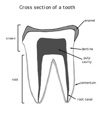
An individual tooth consists of an exposed crown and a root , buried in the gum and jaw. The crown is usually at least partly covered by an outer layer of an especially hard substance related to bone called enamel . Beneath the enamel (and sometimes exposed to the surface if the enamel is missing or worn away) is an intermediate layer of material called dentine , which is also similar to bone but is not nearly as hard as enamel. It surrounds an inner pulp cavity filled with pulp (a living, vascular and well innervated tissue). Blood vessels and nerves reach the pulp cavity through a channel, the root canal , that penetrates the root. An additional layer of bony material, cementum , usually surrounds the root.
As most teeth mature, the root canal gradually closes and the pulp cavity is sealed off. These teeth are called " ." In contrast, " " teeth are those in which the root canal remains open and the tooth continues to grow indefinitely. Rodent incisors and the molars of many arvicoline rodents are examples of rootless or evergrowing teeth; the molars of dogs and humans are rooted.
Teeth are present in most vertebrates (turtles and modern birds are notable exceptions), and in some groups the diversity of teeth rivals that seen in mammals. A significant distinction of mammals, however, is that mammalian teeth are restricted to just three bones, the maxillary and premaxillary of the upper jaw and the dentary of the lower jaw.
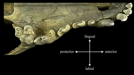
Finally, a note on orientation: mammalogists refer to " labial ," " lingual ," and " occlusal " surfaces. The labial side of the tooth is the side closest to the lips; the lingual side lies next to the tongue. The occlusal surface is the surface that meets a tooth or teeth in the opposite jaw during chewing.

